Breast Cancer and AI: better image quality
Breast Cancer and AI: better image quality as the key to faster diagnosis
Radiologists rely on MRI and mammography images to detect breast cancer. While both are well-established diagnostic tools, there is still room for improvement in terms of accuracy. The Austrian start-up Salataris AI is currently developing an AI model that could help achieve this. One of the first steps is to enhance image quality – a crucial factor for accurate diagnosis. This article explains how the team is approaching the challenge.
Bettina Benesch
Today, the chances of detecting a tumour in the breast are quite high. MRI and mammography are widely and successfully used. However, image quality still presents some challenges. These include noise, low resolution, weak contrast, image artefacts (such as metal interference), and motion blur caused by breathing or movement during scanning. Improving image quality achieves two important outcomes:
1. Improved diagnostic accuracy:
High-quality images allow radiologists to identify subtle tumours and distinguish malignant changes from benign ones – such as cysts or calcifications. Poor contrast or image noise can lead to false-negative or false-positive results. A false negative means a tumour is overlooked; a false positive may indicate a tumour that isn’t actually there – often leading to unnecessary biopsies.
2. More powerful AI models:
AI models are already supporting medical professionals in diagnostics. These models learn to recognise patterns from the image data they’re trained on. If trained with high-quality images, their performance improves significantly.
Noise and artefacts (image distortions) can mislead the model into learning incorrect patterns. Removing noise from an MRI scan allows the model to focus on actual anatomical structures instead of being distracted by random visual interference. So improving image quality benefits both humans and machines. There are two main ways to achieve this: through traditional methods (such as better hardware) and through deep learning – a subfield of artificial intelligence.
Traditional methods for enhancing medical images
Optimised hardware:
In mammography, high-resolution detectors and compression techniques already help reduce tissue overlap and motion blur.
Advanced MRI coil technology and stronger magnetic fields (e.g. 3 Tesla or 7 Tesla scanners) improve the signal-to-noise ratio (SNR). A high SNR means that the useful signal is much stronger than background noise, resulting in clearer, more detailed images.
Image processing techniques:
Noise-reduction filters such as Gaussian or median filters reduce random noise in MRI images.
Other techniques enhance contrast to make abnormalities more visible.
Additional methods address motion artefacts caused by breathing or trembling – or artefacts resulting from metal implants.
While these traditional approaches are effective, they often require manual adjustments and are limited in handling complex noise patterns.
Using deep learning to improve image quality
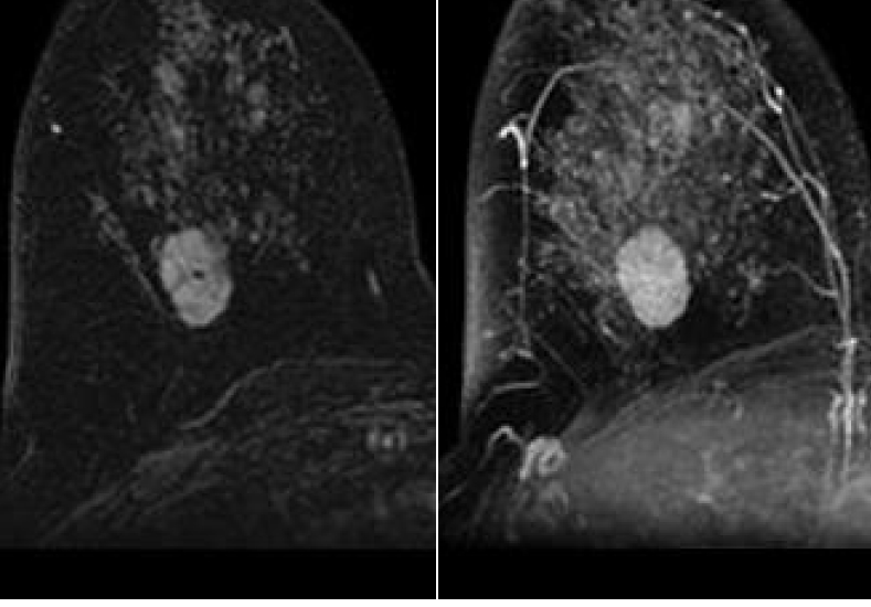
Salataris AI aims to support and enhance these traditional methods with deep-learning technologies. Deep learning involves artificial neural networks that analyse vast amounts of data to independently identify patterns and correlations. Unlike conventional software, these systems aren’t manually programmed but learn directly from the image data. With support from EuroCC Austria, the Salataris AI team trained several models – using both supervised and unsupervised learning – on the Italian supercomputer Leonardo. The goal: better images. All the techniques described here have already been successfully implemented (see examples below).
Deep learning is particularly effective in detecting complex patterns in data, making it ideal for automated and optimised image enhancement. Salataris AI has used a variety of approaches:
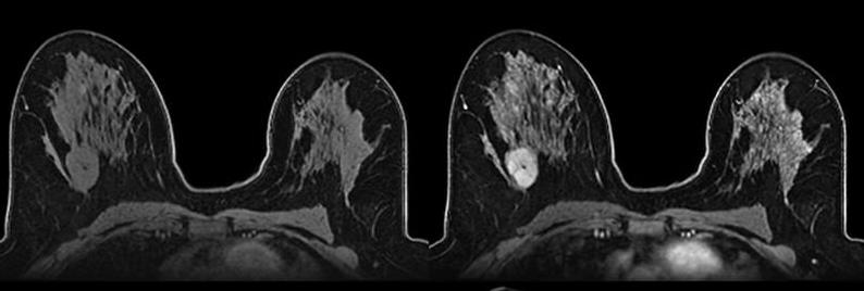
Contrast-enhancing networks
It’s often difficult to differentiate tumours from healthy tissue in mammograms or MRI scans, especially when both appear similar in brightness. That’s where contrast-enhancing networks come in. These special deep learning models analyse the image and detect areas that may be diagnostically important – for example, suspicious structures or tissue changes. They then selectively adjust contrast in those areas, making tumour regions appear brighter or more defined, while surrounding healthy tissue is visually downplayed. The result is clearer images where potential tumours are easier to spot.
Generative Adversarial Networks (GANs)
GANs consist of two competing neural networks – one generates images, the other evaluates them. This “competition” leads to continually improved results, similar to an artist and a critic challenging one another. Salataris AI uses CycleGAN (Cycle-Consistent Generative Adversarial Network) to transform low-quality images into high-quality ones – for instance, sharpening blurry MRI scans. Another tool is SRGAN (Super-Resolution GAN), which enhances the resolution of low-quality mammograms without losing structural detail.
Unsupervised learning to remove artefacts
Unsupervised learning means that the deep learning model does not require predefined “correct” answers – no need for manually labelled images indicating which parts are tumours. Instead, the model identifies patterns by itself and learns to distinguish actual tumour features from noise or artefacts. This is done using convolutional neural networks (CNNs) such as U-Net, which can identify image regions likely to be artefacts and remove them – all without needing paired images (with and without artefacts) for training.
Denoising autoencoders
These neural networks compress noisy images into what’s called a latent space, then reconstruct a cleaner version. An encoder turns the image into a simplified representation (a kind of summary), and a decoder reconstructs an enhanced version of it. This reduces noise in both MRI and mammography images, while retaining important details like tumour boundaries.
Modern breast cancer diagnostics clearly benefit from optimised imaging. The foundation of these improvements is computing power – and Europe’s supercomputers are more than capable of handling the data load.
Free proof-of-concepts on European supercomputers
Deep learning bridges traditional diagnostics and the current demands of personalised, efficient medicine. When enhanced images are combined with tumour recognition algorithms, a positive feedback loop can emerge: better input data leads to more accurate models, enabling radiologists to make faster and more reliable decisions. Modern breast cancer diagnostics clearly benefit from optimised imaging. The foundation of these improvements is computing power – and Europe’s supercomputers are more than capable of handling the data load.
Through EuroCC, proof-of-concept access to supercomputing resources is free for start-ups and SMEs. For more information, please get in touch with our experts directly at: info@eurocc-austria.at.